The transition from acquiring a raw research chemical to executing a flawless cellular assay requires precise technical execution. For young scientists and veteran researchers alike, the physical handling of sensitive peptide chains is a critical step where errors frequently occur. Even the most pure, expensive compounds can be ruined instantly through improper storage, aggressive mixing, or incorrect solvent selection.
When exploring tissue remodeling or collagen pathways, the copper-bound tripeptide requires specific preparation environments to remain completely active. This technical guide outlines the exact laboratory steps needed to transition a newly arrived vial into a usable solution. By standardizing these operational workflows, laboratories can maximize their resources and eliminate costly experimental drift.
Initial Inspection and Thermal Stabilization of Arriving Packages
The moment a sensitive shipment arrives at your receiving dock, the tracking and preservation protocol must begin immediately. Vials should be inspected for structural cracks, loose caps, or signs of moisture intrusion that may have occurred during international transit. Before opening or attempting reconstitution, the vacuum-sealed containers should be allowed to reach room temperature naturally.
Opening a chilled vial in a humid room causes immediate condensation to form inside the glass container, introducing moisture into the dry powder. This unwanted moisture can trigger rapid hydrolysis, breaking the delicate peptide bonds before the compound ever contacts your target cell cultures. Making a smart ghk cu peptide buy decision means nothing if these basic physics principles are ignored during unboxing.
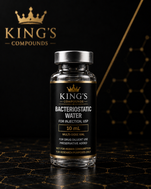
Step-by-Step Reconstitution Protocol for Maximum Stability
To transition a 50mg lyophilized cake into a stable liquid concentrate, researchers must utilize sterile, degassed bacteriostatic water or phosphate-buffered saline. The exact volume added depends on your desired working concentration for the upcoming cellular assays. The following sequence ensures a safe, uniform dissolution process without damaging the underlying molecular structure:
- Sanitize the rubber stopper of the vial using a fresh, single-use seventy percent isopropyl alcohol swab.
- Draw the exact calculated volume of sterile diluent into a high-precision laboratory syringe.
- Angle the needle carefully so the fluid streams slowly down the inner glass wall of the vial.
- Allow the liquid to absorb into the lyophilized cake naturally for two minutes without moving the vial.
- Gently roll the vial between your palms until the solution is completely clear and free of particulate.
Optimal Storage Dispositions for Prepared Aliquots
Once reconstituted, the liquid solution is significantly more vulnerable to thermal degradation and structural breakdown. Researchers should divide the master solution into small, single-use working aliquots using sterile polypropylene tubes to avoid repeated freeze-thaw cycles. These prepared tubes must be stored in a calibrated medical freezer at minus twenty degrees Celsius for long-term stability preservation.
Maximizing Data Accuracy with Premium GHK Cu Peptide in Cellular Assays
With a perfectly prepared solution in hand, researchers can begin introducing the compound into their experimental tissue models. The presence of the copper tripeptide triggers rapid adjustments in cellular migration, cell signaling, and protein secretion rates. Utilizing highly stable ghk cu peptide ensures that the changes observed are purely a result of the molecule's unique chemical profile.
In vitro tracking models demonstrate that the compound helps cells maintain their structural orientation and adhesion qualities under high-stress environments. It acts as a powerful coordinator, ensuring that various cell types work in complete harmony to rebuild damaged extracellular matrix networks. This coordinated action is highly useful for studying vascular development and complex tissue engineering.
Overcoming Common Interference Factors in Peptide Testing
Peptides can sometimes exhibit non-specific binding to standard plastic laboratory equipment, reducing the active concentration available to your cell cultures. To prevent this hidden loss of material, scientists should utilize low-binding plastics or treat surfaces with a mild blocking buffer. This simple procedural adjustment ensures that your cellular models receive the exact calculated dosage intended by your protocol.
Conclusion
The journey to scientific discovery requires absolute precision across every phase of chemical procurement, preparation, and live application. Mastering the technical art of peptide reconstitution guarantees that your testing models remain accurate and highly reliable. By protecting your molecular compounds from environmental trauma, you safeguard your laboratory's data integrity and accelerate paths toward incredible medical insights.